Red/Green Assay
Slides were blocked with 10% fetal bovine
serum for 30 min at RT and subsequently exposed for 60 min to rabbit
anti-P30 (SAG1) (1:1000). Cells were then permeabilized for 10 min in
1% Triton X-100. Monoclonal antibody G11-9 (anti-SAG, 1:500) was added
and incubated at RT for 60 min.
Cells were then permeabilized
for 10 min in 1% Triton X-100. Monoclonal antibody G11-9 (anti-SAG,
1:500) was added and incubated at RT for 60 min. Slides were
subsequently stained with a mixture of goat anti-rabbit IgG (1:500)
conjugated to Alexa 594 (Red) fluorophores, goat anti-mouse IgG (1:500)
conjugated Alexa 488 fluorophores (green) for 60 min at RT.
Slides
were rinsed, mounted and images were captured using a Nikon Eclipse 800
Immunofluorescence microscope with a SPOT RT CCD camera at 600X magnification.
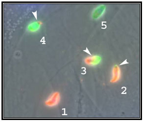
Tachyzoites in Different States of Invasion:
1, Attached, 2-4 Penetrating, 5 Invaded.
Arrow heads indicate site of host cell plasma
membrane penetration.